Cell Analytics

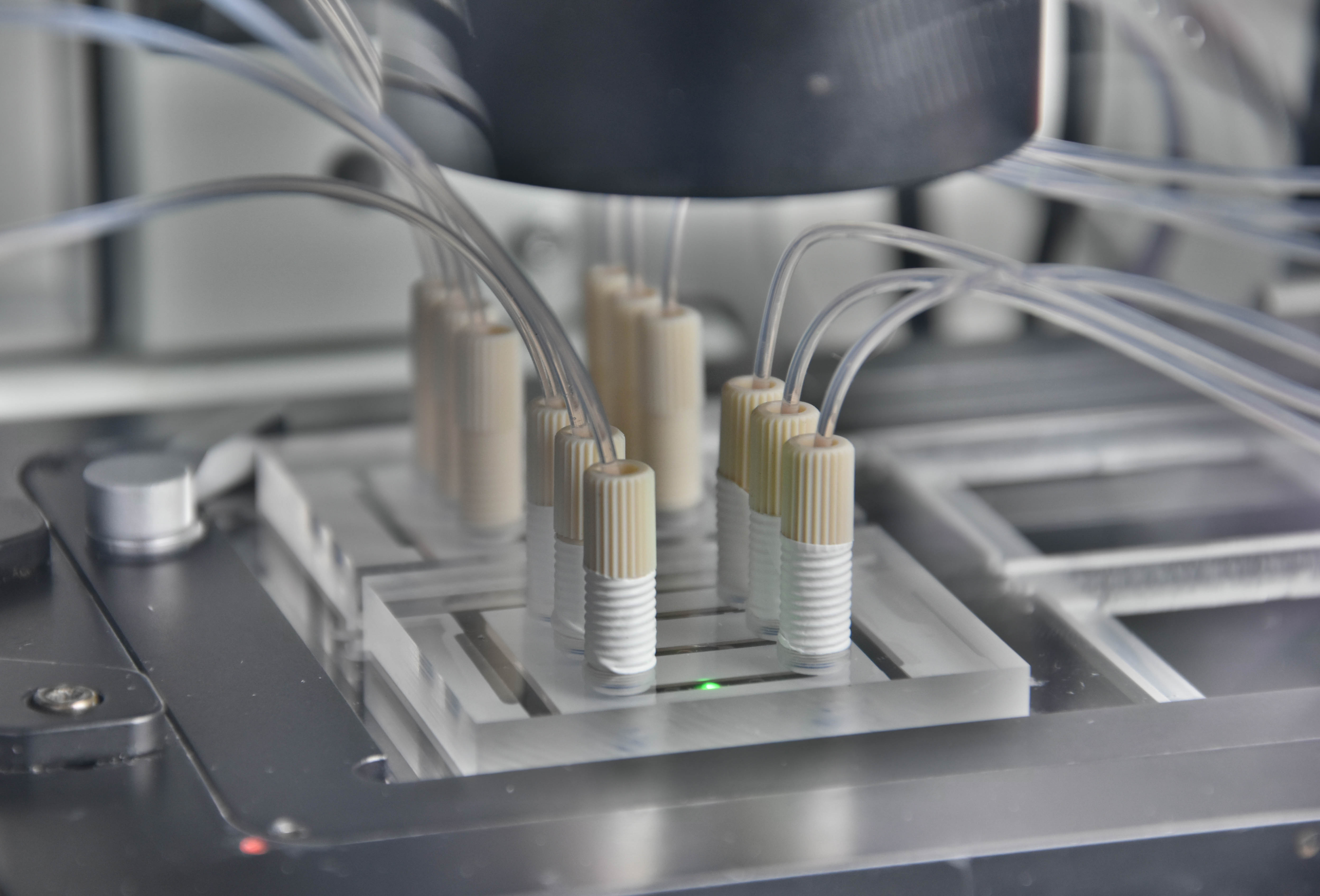
Organ-On-Chip-System: physiologische und kontrollierte Zellkultur in Kombination mit Echtzeitmessungen der Zellvitalität.
© Fraunhofer IZI-BB
Organ-On-Chip-System: physiologische und kontrollierte Zellkultur in Kombination mit Echtzeitmessungen der Zellvitalität.

High-performance methods for the analysis of cells enable an always deeper insight into cell biological processes and thus form the basis for many medical and biotechnological applications. A wide range of methods for the cultivation and analysis of primary cells and cell lines is available at the institute. Cell culture laboratories equipped with state-of-the-art devices and microscopes of safety level S1 or S2 allow working with blood and many other biological cell samples. We develop customized procedures according to your individual requirements and specifications and support you in your preclinical research projects as well as drug or medical device testing.

Range of services

  • Design and development of microbioreactors for long-term cultivation of sophisticated cell models.
  • Development of microfluidic systems for sorting cells and other microparticles
  • Automated image processing: characterization and sorting of cells based on their microscopic image or on morphological markers
  • Integration of microsensors into microfluidic systems for real-time detection of cell media parameters (e.g. oxygen, pH, glucose)
  • Improvement of biocompatibility of synthetic surfaces by coating with polymers and biomolecules and by thermoresponsive polymers for gentle cell detachment
  • Competent processing and manipulation of single cells and smallest cell samples in microfluidic systems
  • Establishment of functional cell assays (e.g. proliferation, cytotoxicity, chemotaxis, neurite growth, stem cell differentiation, intracellular calcium, intracellular pH, etc.)
  • High-end optical microscopy e.g. high-sensitivity fluorescence measurements, time-resolved fluorescence microscopy of living cell systems, near-surface microscopy (TIRF), confocal laser scanning microscopy (LSM)
  • Cell cultivation, cell expansion and transfection service

Real-time sensor technology

© Fraunhofer IZI-BB
Cross-section of a microchannel with six microcavities; embedded with hepatocytes (gray) and optical sensor particles (red: O2, blue: pH) in 3D collagen matrices.

One difficulty in assessing toxicity measurements of new substances such as pharmaceuticals, cosmetics or chemicals/REACH (European Chemicals Regulation on Registration, Evaluation, Authorization and Restriction of Chemicals) is the investigation of meaningful measurands that provide information on the state of the cell tissue. Currently, mainly endpoint analyses (cell staining, gene, protein expression) are performed, which have to be individually adapted to the respective questions. A more generally applicable alternative is based on the measurement of metabolic processes of the cell tissue. By integrating sensors into microfluidic systems for real-time analysis of cell viability, changes in cell metabolism are brought into the temporal context of changes in cultivation conditions, e.g., drug addition and removal. This allows a much more comprehensive elucidation of the mechanisms of action of specific groups of substances and the investigation of repeated doses and combination treatments. For example, cell respiration and pH changes in the immediate vicinity of the cells as well as glucose and lactate concentrations in the cell medium are recorded.

Analysis of chemotaxis in microfluidic systems

Analysis of chemotaxis in microfluidic systems
© Fraunhofer IZI-BB
Phase contrast image of the migration of HFF1 cells in concentration gradients with the corresponding trajectories plotted. Using optical fluorescence methods, changes to the cytoskeleton during the formation of lamellipodia can be monitored dynamically. Scale bar: 50 µm.

Chemotaxis refers to the directed migration of cells in the concentration gradient of a stimulating substance and plays a central role in many important biological processes such as embryonic development, immune defense or cancer. Understanding the underlying molecular mechanisms is essential for developing new drugs targeting these processes. Unfortunately, classical chemotaxis assays often fail to adequately control the experimental framework for studying chemotaxis. To overcome these limitations, we use microfluidic systems that can generate highly stable concentration gradients and dynamically record the migration of stimulated cells over hours. Here, the gradients are generated under laminar flow conditions by diffusion between two parallel partial flows. By varying flow rates, the steepness of the gradient can be adjusted and it can be positioned at different positions in the microfluidic channel. This allows the stimulation of cells with soluble factors to be varied very flexibly in terms of location and time and adapted to the respective experimental conditions.

Calcium signals in single cells

Calcium Signals in Single Cells
© Fraunhofer IZI-BB
Creating a specific contact on one individual T-cell using a functionalized microparticle in a microchannel. The increase in the intracellular Ca2+ concentration triggered by binding the cell to the particle surface was measured using a calcium-sensitive fluorescent dye.

The interaction of cells with their environment is a powerful mechanism to control cellular states in vivo. To decipher the signal transduction processes involved in this process, specific events along the cellular signaling cascade need to be detected and their interrelationships elucidated. This cannot be done by ensemble measurements, since averaging biological data always disregards the variability of the response behavior of individual cells and thus provides blurred results. Only multiparameter analysis at the single cell level can provide the crucial information that is essential for a detailed understanding of cellular signaling pathways. In our group, we are developing techniques and procedures to specifically manipulate the microenvironment of individual cells, allowing soluble or surface-bound stimuli to be presented to the cells individually and in a highly controlled manner. The cellular signal transduction processes induced by these stimuli are analyzed at different temporal scales. In this way, important biological processes such as intercellular communication or the differentiation of cells can be studied very precisely and under reproducible environmental conditions.

Analysis of differentiation processes and "artificial stem cell niches"

stem cells
© Fraunhofer IZI-BB
Stem cells differentiated into a neuronal phenotype in a microfluidic system. Extensions of neural cells spread out from a dense cell cluster radially. Detection of neuronal cells by means of β-III-tubulin (green) and actin (red).

As we know today, the differentiation of stem cells in the organism is strongly controlled by their microenvironment. Other cell types as well as extracellular proteins play a central role in this process. Via changes in this so-called "niche", the stem cell is given a signal, e.g. to rest, to proliferate or to differentiate.

Unfortunately, observing such dependencies is technically difficult, since one does not work with a flat layer of cells in culture, but in the nascent embryo or in the adult organism one has to try to follow, microscope, stain and manipulate individual cells without exerting an undesirable influence on them. This is sometimes done on simple organisms such as C. elegans (nematode) and D. melanogaster (fruit fly). However, on more complex systems, especially in humans, one faces insurmountable difficulties.

We are developing approaches to artificially recreate the "niche" of stem cells and thus export them under a microscope. This will make it possible to study the behavior of individual stem cells in their physiological environment and to gain valuable insights into basic principles such as ways to influence stem cell differentiation. A distant goal is the controlled differentiation and preparation of single cells in chip format.

  • Establishment of functional cell assays (e.g. proliferation, cytotoxicity, chemotaxis, neurite growth, stem cell differentiation, intracellular calcium, intracellular pH, etc.).
  • Optical microscopy at high end level (e.g. high light sensitive fluorescence measurements, time resolved fluorescence microscopy of living cell systems, etc...)
  • Expression analysis by immunostaining and Western blots
  • Detection and quantification of cellular and proteingenic blood components
  • Confocal laser scanning microscope (Zeiss LSM510)
  • Fully automated fluorescence microscopes with climate control for long-term observation of living cells (cellSens, Olympus)
  • Transmitted and reflected light microscopy with bright field, phase contrast, fluorescence, polarization and total internal reflection (TIRFM)
  • Systems for real-time measurement of oxygen consumption (Opal, Colibri) and pH changes near cells

  • Flechner M, Schaller J, Stahl M, Achberger K, Gerike S, Hannappel Y, Fu J, Jaeger M, Hellweg T, Duschl C, Uhlig K. Adhesion, proliferation and detachment of various cell types on thermoresponsive microgel coatings. Biotechnol Bioeng. (2022), 1– 12.
  • Gehre C, Flechner M, Kammerer S, Küpper J-H, Coleman C D, Püschel G P, Uhlig K, Duschl C. Real time monitoring of oxygen uptake of hepatocytes in a microreactor using optical microsensors. Sci Rep (2020) 10, 13700.
  • Bavli D, Prill P, Ezra E, Levy G, Cohen M, Vinken M, Vanfleteren J, Jaeger MS, Nahmias Y. Real-time monitoring of metabolic function in liver-on-chip microdevices tracks the dynamics of mitochondrial dysfunction. PNAS (2016) 113, S. E2231-E2240.
  • Prill S, Bavli D, Jaeger MS, Schmälzlin E, Levy G, Schwarz M, Duschl C, Ezra E, Nahmias Y. A Real-Time Monitoring of Oxygen Uptake in Hepatic Microwell Bioreactor Reveals CYP450-Independent Direct Mitochondrial Toxicity of Acetaminophen multilayers. Archives of Toxicology, 90 (2016) 1181-1191. DOI dx.doi.org/10.1007/s00204-015-1537-2
  • Renner A, Jaeger MS, Lankenau A, Duschl C. Position-dependent chemotactic response of slowly migrating cells in sigmoidal concentration profiles. Appl Phys A. (2013), 112(3), 637-645.
  • Uhlig K, Wischerhoff E, Lutz JF, Laschewsky A, Jaeger MS, Lankenau A, Duschl C. Monitoring cell detachment on PEG-based thermoresponsive surfaces using TIRF microscopy. Soft Matter. (2010), 6, 4262-4267.
  • Habaza M, Kirschbaum M, Guernth-Marschner C, Dardikman G, Barnea I, Korenstein R, Duschl C, Shaked NT. Rapid 3D Refractive-Index Imaging of Live Cells in Suspension without Labeling Using Dielectrophoretic Cell Rotation. Adv. Sci. (2017), 4, 1600205
  • Kirschbaum M, Jaeger MS, Duschl C. Measurement of surface-mediated Ca2+ transients on the single-cell level in a microfluidic Lab-on-a-Chip environment. Methods Mol Biol. (2015);1272:247-56
  • Schreml S, Meier RJ, Kirschbaum M et al. Luminescent Dual Sensors Reveal Extracellular pH-Gradients and Hypoxia on Chronic Wounds That Disrupt Epidermal Repair. Theranostics. (2014), 4, S. 721-735.
  • Renner A, Jaeger MS, Lankenau A, Duschl C. Position-dependent chemotactic response of slowly migrating cells in sigmoidal concentration profiles. Appl Phys A. (2013), 112(3), 637-645.
  • Kirschbaum M, Jaeger MS, Duschl C. Correlating short-term Ca2+ responses with long-term protein expression after activation of single T cells. Lab Chip. (2009), 9, 3517-3525.
  • Kirschbaum M, Jaeger MS, Schenkel T, Breinig T, Meyerhans A, Duschl C. T cell activation on a single-cell level in dielectrophoresis-based microfluidic devices. J Chromatogr A. (2008), 1202, 83–89.

  • Tel Aviv University, OMNI Group
  • Universitätsklinikum Regensburg
  • Universitätsklinikum Tübingen
  • Surflay Nanotec GmbH
  • Colibri Photonics GmbH
Zellanalytik
© Fraunhofer IZI-BB